Multimodal Preclinical Imaging Brings Science Closer to Personalized Medicine
Multimodal Preclinical Imaging Brings Science Closer to Personalized Medicine.
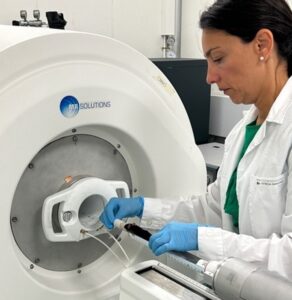
The IIBM-CSIC-UAM has installed state-of-the-art technology that combines magnetic resonance imaging and positron emission tomography.
Did you see the article from the Spanish CSIC?
This is a great read highlighting how using MR Solutions flexible technology in preclinical studies can have a more direct and faster application in clinical practice.
‘One of the main advantages of the new MRI/PET system is the obtaining of greater functional and metabolic information both from a tumor and from the surrounding tissues, by using the multimodal image acquired simultaneously it is possible to detect in real time the accumulation of nanoparticles in the tumor that contain radioisotopes (visible by PET) and contrast agents in magnetic resonance imaging (visible by MRI). This represents a significant advance in the ability of researchers to monitor and assess tumour progression and response to treatment in preclinical models, with important implications for the development of more effective and personalised therapies.
Another undeniable advantage of the equipment is the ability to modify the magnetic field in which it operates, operating at both 3T and 7T. “This allows MRI images to be acquired in both fields: at 7T the image obtained has a higher resolution thanks to the better signal-to-noise ratio, but being able to operate at 3T is essential since it is a field used in hospital facilities,” adds the researcher. This flexibility means that preclinical studies carried out with the new equipment have a more direct and faster application in clinical practice.’
Thanks to Pilar López-Larrubia, Nuria Arias Ramos and Teresa Navarro Hernanz for this publication.
Read the full article here.