
Anatomical Imaging
Anatomical imaging has revolutionized medical science. It has been estimated that during the past 20 years there has been as much progress in clinical medicine as in all the previous history of medicines combined, and anatomical imaging has made a major contribution to that progress. Anatomical imaging allows medical personnel to look inside the body with amazing accuracy and without the trauma and risk of exploratory surgery. Although most of the technology of anatomical imaging is very new, the concept and earliest technology are quite old.
MRI provides information to qualitatively and quantitatively describe the shape, size, and integrity of gray and white matter structures in the brain. Morphometric techniques measure the volume or shape of gray matter structures, such as subcortical nuclei or the hippocampus, and the volume, thickness, or surface area of the cerebral neocortex. Macrostructural white matter integrity can also be measured using volumes of normal and abnormal white matter, providing indications of inflammation, edema, or demyelination, complementing microstructural diffusion weighted MRI to provide a comprehensive picture of white matter integrity. Since brain function depends to some extent on the integrity of brain structure, measures that characterize the underlying tissue integrity also allow one to examine the impact of tissue loss or damage on functional signals. Furthermore, structural MRI provides an anatomical reference for visualization of activation patterns and regions of interest to extract functional signal information.
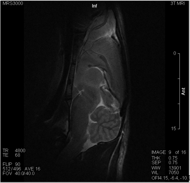
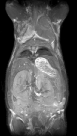 MRI may be used to examine bones, joints, and soft tissues such as cartilage, muscles, and tendons for injuries or the presence of structural abnormalities or certain other conditions, such as tumors, inflammatory disease, congenital abnormalities, osteonecrosis, bone marrow disease, and herniation or degeneration of discs of the spinal cord. MRI may be used to assess the results of corrective orthopedic procedures.
References:
http://www.mhhe.com/biosci/ap/saladin/humbody/reading1.mhtml
http://fmri.ucsd.edu/Howto/3T/structure.html
http://www.hopkinsmedicine.org/healthlibrary/test_procedures/orthopaedic/magnetic_resonance_imaging_mri_of_the_bones_joints_and_soft_tissues_92,P07652/