
Neurology
 Brain imaging falls into two broad categories:
Structural imaging, which deals with the structure of the brain and the diagnosis of large-scale intracranial disease (such as tumor), and injury.
Functional imaging, which is used to diagnose metabolic diseases and lesions on a finer scale.
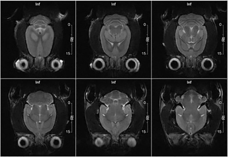
MRI Advantage
In some cases, MRI can provide clear images of parts of the brain that can’t be seen as well with an X-ray, CAT scan, or ultrasound, making it particularly valuable for diagnosing problems with the pituitary gland and brain stem. The main advantage of the MRI over other techniques is the tissue contrast, and it has fewer artifacts than CT e.g. when viewing the brainstem.
Functional MRI
Functional magnetic resonance imaging, or fMRI, is a technique for measuring brain activity. It works by detecting the changes in blood oxygenation and flow that occur in response to neural activity – when a brain area is more active it consumes more oxygen and to meet this increased demand blood flow increases to the active area. fMRI can be used to produce activation maps showing which parts of the brain are involved in a particular mental process.
Ideal technique for:
- Blood flow or blood clots to the brain. MRI can show bleeding in or around the brain
- Symptoms of a known or suspected head injury
- Symptoms such as change in consciousness, confusion, or abnormal movements. These symptoms may be caused by brain diseases, such as Huntington’s disease, multiple sclerosis Parkinson’s disease, or Alzheimer’s disease.
- Hydrocephaly
- Tumors, infections, an abscess, or conditions of the brain or brain stem, such as encephalitis or meningitis
- Optic nerves and auditory nerves
- Alterations of the pituitary gland
References:
http://en.wikipedia.org/wiki/Magnetic_resonance_imaging_of_the_brain http://www.ndcn.ox.ac.uk/departments/FMRIB/research/introduction-to-fmri