
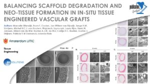
BALANCING SCAFFOLD DEGRADATION AND NEO-TISSUE FORMATION IN IN-SITU TISSUE ENGINEERED VASCULAR GRAFTS
Congratulations to the authors of this week’s publication spotlight!
Marcelle Uiterwijk, Bram F. Coolen, Jan-Willem van Rijswijk, Serge H.M. Söntjens, Michel H.C.J. van Houtem, Wojciech. Szymczyk, Laura Rijns, Henk M. Janssen, Allard van de Wal, Bouten A.J.M. de Mol, Carlijn.V.C. Bouten, Gustav J. Strijkers, Patricia Y.W Dankers & Jolanda Kluin
An essential aspect of cardiovascular in situ tissue engineering (TE) is to ensure balance between scaffold degradation and neo-tissue formation. We evaluated the rate of degradation and neo-tissue formation of three electrospun supramolecular bisurea-based biodegradable scaffolds that differ in their soft-block backbone compositions only. Scaffolds were implanted as interposition grafts in the abdominal aorta in rats, and evaluated at different time points (t = 1, 6, 12, 24 and 40 weeks) on function, tissue formation, strength and scaffold degradation. The fully carbonate-based biomaterial showed minor degradation after 40 weeks in vivo, while the other two ester-containing biomaterials showed (near) complete degradation within 6 to 12 weeks. Local dilatation was only observed in these faster degrading scaffolds. All materials showed to some extent mineralization, at early as well as late time points. Histological evaluation showed equal and non-native like neo-tissue formation after total degradation. The fully carbonate based scaffolds lagged in neo-tissue formation, presumably as its degradation was (far from) complete at 40 weeks. A significant difference in vessel wall contrast enhancement was observed by MRI between grafts with total compared to minimal degraded scaffolds.
The data was acquired on a cryogen-free MRS*DRYMAG 7T system at The Amsterdam University Medical Center. The article was published in Tissue Engineering in 2024.
The publication is available at: 10.1089/ten.TEA.2023.0019