
Oncology
Functional MRI
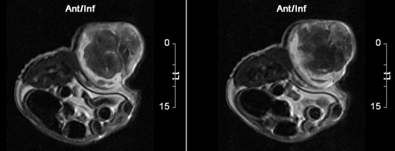
Functional magnetic resonance imaging is rapidly evolving as a capable non-invasive assessment tool for oncology to improve diagnosis and to monitor therapy. Current clinical techniques are based on microcirculation imaging using extracellular low molecular weight contrast agents such as gadopentetate dimeglumine and analogues. The temporal evolution of the enhancement visualizes the angiogenic properties of lesions with regard to vascular density and permeability, heterogeneity, and changes during therapy.
Ideal technique for:
- Finding a tumor.
- Determine tumor nature.
- Evaluate stage of cancer (size and location of the tumor).
- Plan treatments, surgery or radiation therapy.
- Monitor tumor’s response to treatment.
- Lymph node evaluation.
- Tumour angiogenesis.
- Suspected recurrent disease.
References: http://mct.aacrjournals.org/content/2/4/419.full