
Angiography
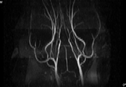
A magnetic resonance angiogram (MRA) is a type of magnetic resonance imaging (MRI) scan that uses a magnetic field and pulses of radio wave energy to provide pictures of blood vessels inside the body. In many cases MRA can provide information that can’t be obtained from an X-ray, ultrasound, or computed tomography.
MRA can find problems with the blood vessels that may be causing reduced blood flow. The condition of the blood vessel walls can also be seen. The test is often used to look at the blood vessels that go to the brain, kidneys, and legs. Information from an MRA can be saved and stored on a computer for further study.
Ideal technique for:
- Aneurysms or weakness in the wall of an artery
- Narrowing of the aorta, or aortic coarctation
- Bleeding in and along the wall of the aorta, or aortic dissection
- Evidence of stroke
- Signs of heart disease
- Narrowing or blockage of the vessels in the arms or legs
- Renal artery stenosis, a narrowing of the blood vessels in the kidneys that can lead to high blood pressure and even renal failure
References:
http://www.hopkinsmedicine.org/healthlibrary/test_procedures/cardiovascular/magnetic_resonance_angiography_mra_135,14/
http://www.webmd.com/heart-disease/magnetic-resonance-angiogram-mra